Atlas of Fundus Autofluorescence Imaging
-
- Hardcover
- Taschenbuch ausgewählt
- eBook
-
Sprache:Englisch
128,99 €
inkl. gesetzl. MwSt.,
Beschreibung
Produktdetails
Einband
Taschenbuch
Erscheinungsdatum
15.10.2010
Abbildungen
XIII, 200 illus. in color., farbige Illustrationen
Herausgeber
Frank G. Holz + weitereVerlag
Springer BerlinSeitenzahl
341
Maße (L/B/H)
24/16,8/1,9 cm
Gewicht
677 g
Auflage
Softcover reprint of hardcover 1st ed. 2007
Sprache
Englisch
ISBN
978-3-642-09119-3
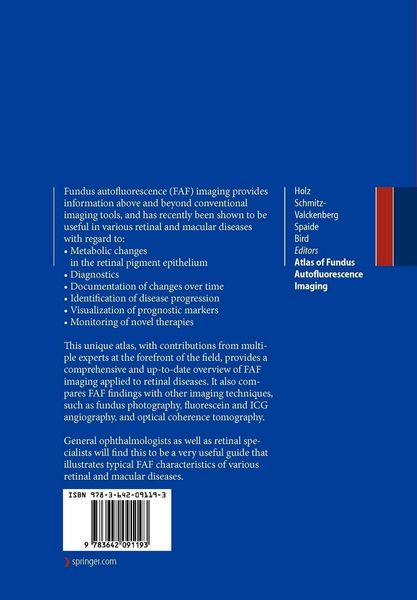
During recent years, FAF (Fundus autofluorescence) imaging has been shown to be useful in various retinal diseases with regard to diagnostics, documentation of changes, identification of disease progression, and monitoring of novel therapies. Hereby, FAF imaging gives additional information above and beyond conventional imaging tools.
This unique atlas provides a comprehensive and up-to-date overview of FAF imaging in retinal diseases. It also compares FAF findings with other imaging techniques such as
fundus photograph, fluorescein- and ICG angiography as well as optical coherence tomography.
General ophthalmologists as well as retina specialists will find this a very useful guide which illustrates typical FAF characteristics of various retinal diseases.
Noch keine Bewertungen vorhanden
Verfassen Sie die erste Bewertung zu diesem Artikel
Helfen Sie anderen Kundinnen und Kunden durch Ihre Meinung.
Kurze Frage zu unserer Seite
Vielen Dank für Ihr Feedback
Wir nutzen Ihr Feedback, um unsere Produktseiten zu verbessern. Bitte haben Sie Verständnis, dass wir Ihnen keine Rückmeldung geben können. Falls Sie Kontakt mit uns aufnehmen möchten, können Sie sich aber gerne an unseren Kund*innenservice wenden.
zum Kundenservice