Analytical Transmission Electron Microscopy An Introduction for Operators
-
- Hardcover
- Taschenbuch ausgewählt
- eBook
-
Sprache:Englisch
48,99 €
inkl. gesetzl. MwSt.,
Beschreibung
Produktdetails
Einband
Taschenbuch
Erscheinungsdatum
23.08.2016
Abbildungen
XVII, 238 illus., 33 illus. in color., farbige Illustrationen, schwarz-weiss Illustrationen
Verlag
Springer NetherlandSeitenzahl
348
Maße (L/B/H)
23,5/15,5/2 cm
Gewicht
557 g
Auflage
Softcover reprint of the original 1st edition 2014
Sprache
Englisch
ISBN
978-94-017-7988-3
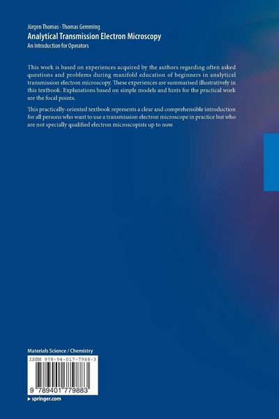
This work is based on experiences acquired by the authors regarding often asked questions and problems during manifold education of beginners in analytical transmission electron microscopy. These experiences are summarised illustratively in this textbook. Explanations based on simple models and hints for the practical work are the focal points.
This practically- oriented textbook represents a clear and comprehensible introduction for all persons who want to use a transmission electron microscope in practice but who are not specially qualified electron microscopists up to now.
Noch keine Bewertungen vorhanden
Verfassen Sie die erste Bewertung zu diesem Artikel
Helfen Sie anderen Kundinnen und Kunden durch Ihre Meinung.
Kurze Frage zu unserer Seite
Vielen Dank für Ihr Feedback
Wir nutzen Ihr Feedback, um unsere Produktseiten zu verbessern. Bitte haben Sie Verständnis, dass wir Ihnen keine Rückmeldung geben können. Falls Sie Kontakt mit uns aufnehmen möchten, können Sie sich aber gerne an unseren Kund*innenservice wenden.
zum Kundenservice